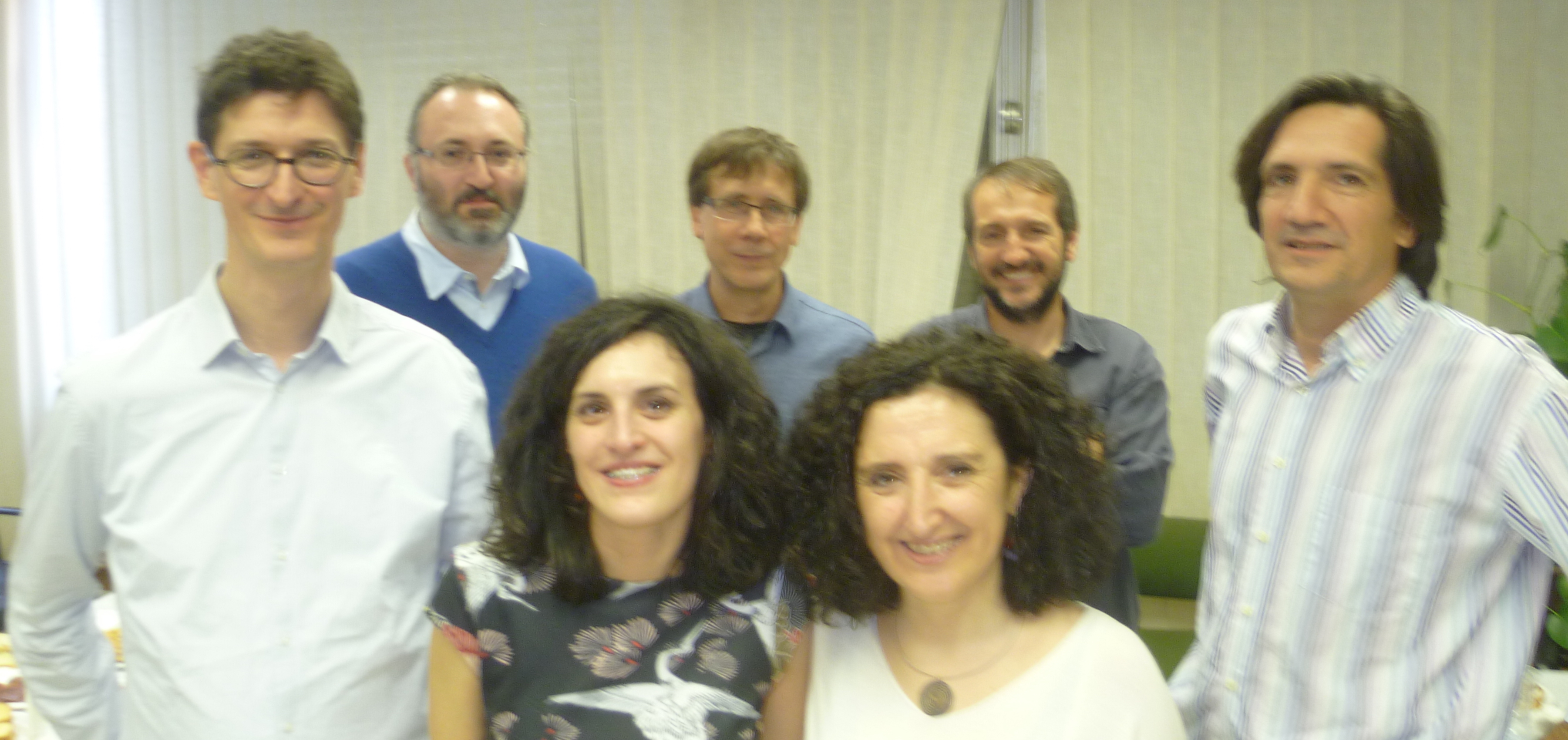
Factuel part à la rencontre des premiers doctorants recrutés dans le cadre de l’initiative « Lorraine Université d’Excellence ». Rencontre avec Mélanie Roland qui effectue sa thèse au laboratoire IAM (et au sein de l’école doctorale RP2E) sous la direction de Nicolas Rouhier et Jérémy Couturier.
Monthly Archives: March 2017
Article: Scientific reports
Populus trichocarpa encodes small, effector-like secreted proteins that are highly induced during mutualistic symbiosis JM Plett, H Yin, R Mewalal, R Hu, T Li, P Ranjan, S Jawdy, HC De Paoli, … Scientific Reports 7 (1), 382
Abstract
During symbiosis, organisms use a range of metabolic and protein-based signals to communicate. Of these protein signals, one class is defined as ‘effectors’, i.e., small secreted proteins (SSPs) that cause phenotypical and physiological changes in another organism. To date, protein-based effectors have been described in aphids, nematodes, fungi and bacteria. Using RNA sequencing of Populus trichocarpa roots in mutualistic symbiosis with the ectomycorrhizal fungus Laccaria bicolor, we sought to determine if host plants also contain genes encoding effector-like proteins. We identified 417 plant-encoded putative SSPs that were significantly regulated during this interaction, including 161 SSPs specific to P. trichocarpa and 15 SSPs exhibiting expansion in Populus and closely related lineages. We demonstrate that a subset of these SSPs can enter L. bicolor hyphae, localize to the nucleus and affect hyphal growth and morphology. We conclude that plants encode proteins that appear to function as effector proteins that may regulate symbiotic associations.
Redox meeting and Conversation
A redox meeting will be held the 29-31 March in Amphitheater 8, Faculté des Sciences, Boulevard des Aiguillettes, Vandoeuvre.
Program redox meeting Nancy March 2017 FINAL
A paper entitled “Redox Regulation : Historical Background and Future Perspectives”
http://theconversation.com/biologie-retour-sur-lhistoire-de-la-regulation-redox-74656
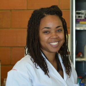
Newcomer: Elodie Sylvestre Gonon
| Name | SYLVESTRE GONON Elodie elodie.sylvestre-gonon9@etu.univ-lorraine.fr |
 |
| Team | Stress response and redox regulation team | |
| Supervisors | A. Hecker / N. Rouhier | |
| Subject | Biochemical characterization of glutathione transferases potentially involved in coumarin metabolism in Arabidopsis thaliana | |
| Type of study | Internship: Master Student (M2) | |
| Period | January – June, 2017 |
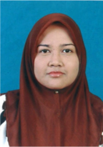
Newcomer: Farah HANANI AZMAN
| Name | HANANI AZMAN Farah farazman7191@gmail.com |
 |
| Team | Stress response and redox regulation team | |
| Supervisor | E. Gelhaye | |
| Subject | Production and purification of protein from aquilaria | |
| Type of visit | Master exchange coming from University Putra – Malaysia | |
| Period | March – May, 2017 |
Newcomer: Liang Li
| Name | LI Liang liang.li@cragenomica.es |
 |
| Team | Stress response and redox regulation team | |
| Supervisors | N. Rouhier/ J. Couturier | |
| Subject | Biochemical analysis of the redox control of aggregation of the plant metacaspase AtMC1 | |
| Type of visit | PhD exchange coming from the CRAG of Barcelona | |
| Period | February to May, 2017 |
New postdoctoral researcher: Shingo MIYAUCHI
| Name | MIYAUCHI Shingo shingo.miyauchi@inra.fr |
 |
| Team | Ecogenomics of interactions team | |
| Supervisor | F. Martin | |
| Subject | Comparative fungal genomics and transcriptomics, and development of analytical and visualization tools for large-scale biological data | |
| Period | 03/2017 – 05/2017 |
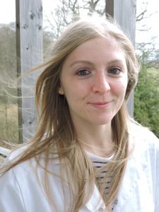
Newcomer: Chloé Viotti
| Name | VIOTTI Chloé chloe.viotti@laposte.net |
 |
| Team | Ecogenomics of interactions team | |
| Supervisor | M. Buée | |
| Subject | Seasonal dynamics of tree N and C reserves (Quercus and Fagus) and monitoring of root microbiota associated functions | |
| Type of study | Internship: Master Student (M1 – 1st year) | |
| Period | February-April, 2017 |
PhD Defense: Leticia Perez
Article: Journal of Visualized Experiments
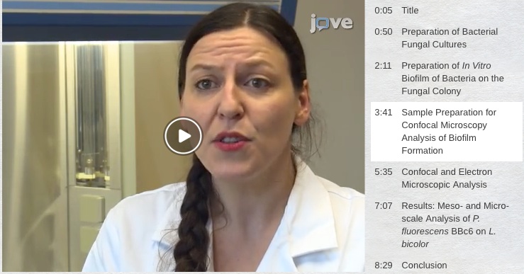
A New Method for Qualitative Multi-scale Analysis of Bacterial Biofilms on Filamentous Fungal Colonies Using Confocal and Electron Microscopy. CM Guennoc, C Rose, F Guinnet, I Miquel, J Labbé, A Deveau. JoVE (Journal of Visualized Experiments), e54771-e54771
Summary
Bacterial biofilms frequently form on fungal surfaces and can be involved in numerous bacterial-fungal interaction processes, such as metabolic cooperation, competition, or predation. The study of biofilms is important in many biological fields, including environmental science, food production, and medicine. However, few studies have focused on such bacterial biofilms, partially due to the difficulty of investigating them. Most of the methods for qualitative and quantitative biofilm analyses described in the literature are only suitable for biofilms forming on abiotic surfaces or on homogeneous and thin biotic surfaces, such as a monolayer of epithelial cells.
While laser scanning confocal microscopy (LSCM) is often used to analyze in situ and in vivo biofilms, this technology becomes very challenging when applied to bacterial biofilms on fungal hyphae, due to the thickness and the three dimensions of the hyphal networks. To overcome this shortcoming, we developed a protocol combining microscopy with a method to limit the accumulation of hyphal layers in fungal colonies. Using this method, we were able to investigate the development of bacterial biofilms on fungal hyphae at multiple scales using both LSCM and scanning electron microscopy (SEM). This report describes the protocol, including microorganism cultures, bacterial biofilm formation conditions, biofilm staining, and LSCM and SEM visualizations.


